Linear Energy Transfer

The density of ionization in particle track is described by the term Linear Energy Transfer (LET). In other words, it is the measure of number of ions produced per unit path length of the track.

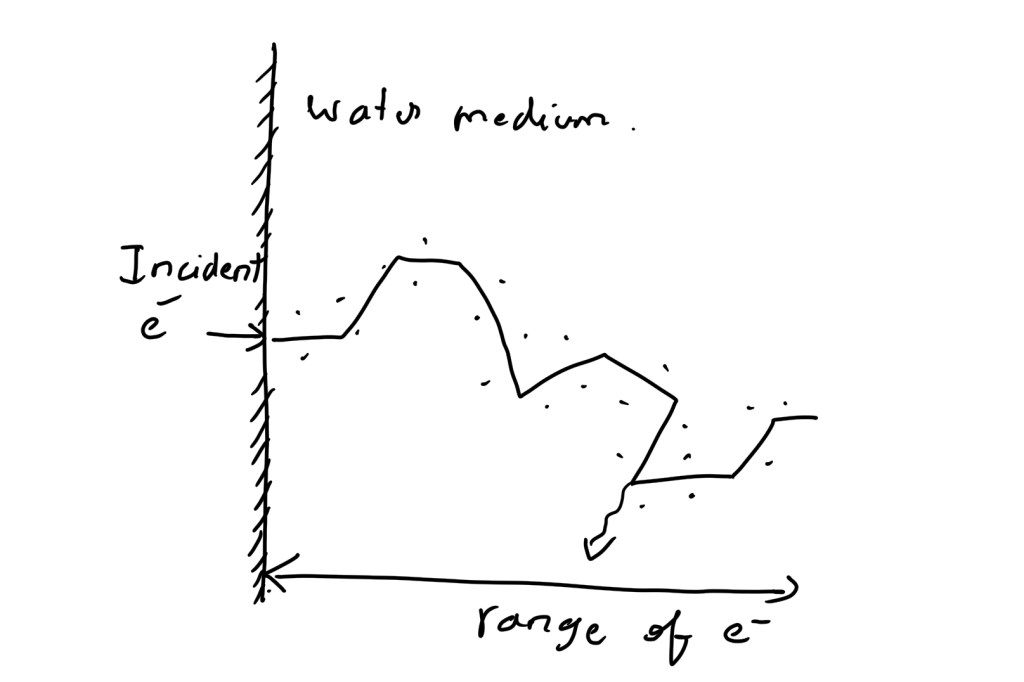
What is difference between path length and range?
The thickness of medium required for entire energy of the particle to be absorbed is called range
The entire zig-zag distance travelled is called path length

Is = Sc/W

Where,
Is is linear density of Ionization.
Sc is unrestricted collision stopping power
W is avg. energy expended per ionization event.

Not all energy accounted for for stopping power Sc results in ionization along the primary track. Instead some secondary electrons will have energy to travel farther away from primary track and deposit the energy. LET Δ is defined to distinguish the ionization event occurring along the primary track from the ionization occurring due to secondary electrons farther away from primary track. The smaller the size of specific energy Δ the stricter the determination of LET Δ with respect to ionization along primary track. The higher the Δ value the LET Δ equals to Sc, which is represented as LET (upper limit of LET).

LET is the average energy per unit distance deposited by charged particle.
Unit KeV /µm

Why LET is an average quantity?
At microscopic level the energy per unit length of track varies drastically.
(one of the complication, If the range of energy variation is too large then the average quantity itself becomes meaning less)

The definition says charged particle, but neutrons are not charged particles? How do we consider them as high- LET radiation?
The charged particles undergoes ionization by interacting with orbital electrons. In case of neutron when they pass through the tissue they do not directly produce ionization. Instead it reacts with the atomic nuclei and eject densely ionizing protons and other particles The ionization due these secondary particles results in high-LET

Different ways to calculate the average?
1] Track Average
2] Energy Average

Track Average – It is obtained by dividing the tracks into equal lengths and calculating the energy deposited in each length. It is most commonly used method
Energy Average – It is obtained by dividing the track into equal energy
intervals and averaging the lengths of the track.

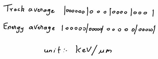
The track average and energy average is almost same for X-rays and mono energetic charged particle. In case of neutron both average shows large variation between them. The biological effects more correlated towards energy average in case of neutron.
e.g. 14 MeV neutron’s – LET track average 12 KeV /µm and energy average 100 KeV /µm

Linear Energy Transfer Values

Radiation TypeLET in KeV /µm
Cobalt – 60 γ rays 0.2
250 KV X-rays 2.0
10 MeV Protons4.7
150 MeV Protons0.5
14 MeV NeutronsTrack Average 12
Energy average 100
2.5 MeV α particle166

It can be simply used to indicate the quality of radiation type(radiobiologically). It is also noted that due to above seen complication such as difference between different averaging methods and range of the average, LET may be misleading in some circumstances.

Reference
1) Hall, Eric J., and Amato J. Giaccia. Radiobiology for the Radiologist. Vol. 6. 2006.
2) Joiner, Michael C., and Albert Van der Kogel. Basic clinical radiobiology fourth edition. CRC press, 2009.
3) Jayaraman, Subramania, and Lawrence H. Lanzl. Clinical radiotherapy physics. Springer Science & Business Media, 2011.

The time scale of effects in radiobiology 🧫

The process are divided into three phases

  1. Physical Phase
  2. Chemical Phase
  3. Biological Phase

Physical Phase

In physical phase the interaction between the charged particle with the atoms of the tissues occurs. It mainly interacts with orbital electrons, ejecting some of them from atoms while exciting some electrons to higher energy levels. The secondary electrons with sufficient energy can induce further ionization and excitation along the tract(cascade of ionization events). In case of indirect action the fast electrons occurs in approximately 10-15 seconds. A high speed electrons takes about 10-18 seconds to traverse the DNA and 10-14 seconds to traverse the mammalian cells. for the volume of every 10 µm for 1 Gy of absorbed radiation dose almost 105 ionization events occur

Incident X-ray photon
⬇️
Fast electron 10-15 seconds

Chemical Phase

Fast electron
⬇️
Ion radical
⬇️
Free Radical
⬇️
chemical changes from breakage of bonds – 1ms(approx.)

About 80% of the cell in composed of water, when the radiation interacts with water molecules H2O \displaystyle \LARGE \rightarrow H2O+ + e . the H2O+ ion radical is formed. this ion radical further reacts with H2O to give H3O+ and OH* where OH* is a free radical. Free radical are highly reactive and they will undergo successive reaction to restore the electronic charge equilibrium. The free radical reactions are completed within 1 ms. The important process of chemical phase is the reactions which inactivate the free radicals e.g. reaction with sulphdryl compounds. Then the process of fixation reaction where the stable chemical changes are induced in biologically significant molecules. Here the fixation means not repair, it means that it made sure that the chemical change or damage is permanent.

Biological Phase

Chemical Changes from the breakage of bond
⬇️
Biological effect (days.months,years,
may not happen within human life span)

The biological effect occurs as the consequence of bonds broken. It begins with the enzymatic reaction that occurs on the residual chemical change. While vast majority of the cells repair, few may lead to cell death.

what is cell death? 
1) loss of specific function - differentiated cells(nerve,muscles,secretory cells) 2) loss of ability to divide - proliferating cells such as stem cells 3) loss of reproductive integrity  

During the first few week and months the loss of stem cells due to the radiation is the early manifestation of normal tissue damage and the early effects are also important for tumours as they are early responding tissues.
The secondary effect of cell killing is cell proliferation, which occurs both in tumours as well as normal tissues. for normal tissue it is an important mechanism as it reduces the acute side effect
The late reactions often occur after years of radiation exposure e.g. spinal cord damage, blood vessels damage and radiation carcinogenesis

what is ion radical?

ion means electrically charged and radical means having unpaired electron in valance shell.
H2O \displaystyle \LARGE \rightarrow H2O+ + e
H2O+ is a ion radical

What is free radical?

Free radicals do not have charge but have unpaired electron in the valance shell.
H2O+ + H2O \displaystyle \LARGE \rightarrow H3O+ + OH*
OH* is the free radical

Reference
Joiner, Michael C., and Albert Van der Kogel. Basic clinical radiobiology fourth edition. CRC press, 2009.

[Most of the sentences written here are taken from basic clinical radio biology book. all credits go the authors of the book mentioned in reference ]